Prof. Edoardo Di Naro
domenica 25 marzo 2018
Musica Rilassante per Bambini | Musica per Addormentarsi, Gravidanza e T...
La mia play list di musica rilassante per mamma e bambino
dott. Edoardo Di Naro Ginecologo a Noicattaro.
giovedì 22 marzo 2018
domenica 11 marzo 2018
https://link.springer.com/article/10.1245%2Fs10434-015-4655-4 (leggi tutto)
- Daniele Bolla
- Sarah In-Albon
- Andrea PapadiaEmail author
- Edoardo Di Naro
- Maria Luisa Gasparri
- Michael M. Mueller
- Luigi Raio
Gynecologic Oncology
First Online: 03 June 2015
- 158Downloads
Abstract
Purpose
The aim of this present study was to evaluate the sonographic correlation between Doppler flow characteristics of the uterine arteries and tumor size in patients with cervical cancer, in order to establish a new potential marker to monitor treatment response.
Methods
This was a retrospective cohort study of 25 patients who underwent a sonographic evaluation of Doppler flow characteristics of the uterine arteries before surgery or radiochemotherapy for early and locally advanced/advanced cervical cancer, respectively, was analyzed. The primary outcome was the correlation between Doppler flow characteristics of the uterine arteries and tumor size in patients with cervical cancer.
Results
Median age was 49 (range 26–85) years, and mean tumor size was 40.8 ± 17 mm. A significant positive correlation was found between tumor diameter and the uterine artery end-diastolic velocity (r = 0.47, p < 0.05) as well as the peak systolic velocity (r = 0.41, p < 0.05). No correlation was found between tumor size and the pulsatility index or resistance index.
Conclusions
In cervical cancer, uterine artery velocity parameters are associated with tumor size. This finding could become particularly useful in the follow-up of locally advanced cervical cancer patients undergoing radiochemotherapy or in corroborating the selection of women with more possibility of a high response rate during neoadjuvant chemotherapy before surgery.
sabato 10 marzo 2018
Prof. Edoardo Di Naro
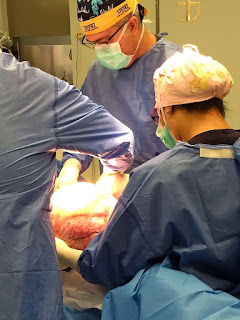
Il Prof. Edoardo Di Naro durante un intervento chirurgico su una paziente di 40 anni.
A 40-year-old woman with dichorionic twins and mitral valve thrombus underwent emergent care when a CT scan and angiography with 3D reconstruction revealed aortal occlusion. This striking image helped to mobilize care by medical personnel from half a dozen departments.
sabato 3 marzo 2018
martedì 27 febbraio 2018
Iscriviti a:
Post (Atom)


